
Photo credits: Dale Ramos
The CZ Biohub Series – Part III
On the third installment of our CZ Biohub series, we visit a lab nicknamed “Le Petit Hotel de Poisson” (The Tiny Fish Hotel) where scientists are using zebrafish as a model organism to study human development. Along the way, we’ll meet Loïc A. Royer, who leads a team at Biohub including Merlin Lange and Shruthi VijayKumar who are using something called a light-sheet microscope to document the incredible complexity of individual cells as they develop into fully functioning organisms.
Manu Leonetti and Sandy Schmid from previous CZ Biohub episodes also join us on the tour.
Watch the videos created with the light sheet microscope below:
In this episode, you will learn the following:
1. Discover how human and fish embryos are surprisingly similar despite their adult forms being vastly different.
2. Uncover how computer vision and algorithms are used to make sense of the transparent, tiny embryos.
3. Unravel the incredible complexity of individual cells and how they communicate to build a functioning organism.


Resources:
Learn more about the zebrafish research being done at the CZ Biohub: https://zebrahub.ds.czbiohub.org/
Learn more about the light sheet microscope at CZ Biohub:
Other episodes from the CZ Biohub Series:
096: Bioengineering Malaria with Paul Lebel
097: Mapping the Cell with Manuel Leonetti
Connect with The Show About Science:
Instagram: https://www.instagram.com/showaboutscience
Facebook: https://www.facebook.com/theshowaboutscience
YouTube: https://www.youtube.com/showaboutscience
Twitter: https://www.twitter.com/natepodcasts
LinkedIn: https://www.linkedin.com/
Loved this episode? Leave us a review and rating wherever you listen to podcasts!
Episode Transcript:
Nate: On the last episode of The Show About Science, we introduced you to Manu Leonetti.
Manu Leonetti: So I’m part of a project at the Biohub called Quantitative Cell Sciences where all of us are really trying to understand how the human body is built, how the human body is organized and we are all asking this kind of question at a different level. I’m interested in zooming in at the level of one single cell and really trying to understand how one cell is organized at the level of all these molecular building blocks that make it, which are the different proteins that make the cell.
Nate: On this episode, we’re going to try to zoom out and look at how the human body is organized at the multicellular level and we’re going to meet three scientists who are trying to understand how a single cell can develop into something like us.
Manu Leonetti: The people you’re going to meet there are trying to understand how different organs are made up of different cells that come together or how an entire developing embryo is made of different organs that are moving together, things like that.
Nate: But you’ll be surprised to learn that the organism these scientists are researching is not us humans. It’s actually a little fish that we’ve done an episode on before. It’s our old friend the zebrafish and this time we’re going to see them in person.
Hello everyone, this is your host Nate and I’m here with the third installment of our series on the Chan Zuckerberg Biohub in San Francisco. We’re here to take a look at the research scientists are doing to cure, prevent, or manage all diseases by the end of the century. So, let’s jump back in and meet our guests today. So, could you introduce yourself? Sure.
Loïc Royer: I’m Loïc Reyer. I’m a group leader here at the Biohub and I lead a team that’s trying to understand how embryos develop.
Nate: Loïc and his team work with something called the light sheet microscope, which we’ll talk about later in the show. They use this specialized microscope to create images and videos of developing embryos.
Loïc Royer: And we also use for that a combination of software and algorithms for computer vision to be able to make sense of these images. And we use all these technologies to try to understand how you build an organism, how you have an embryo that develops from a single cell to a fully functional organism.
Nate: I think that you have one of the coolest jobs in the world.
Loïc Royer: I would agree with that. And I have also the coolest team in the world.
Nate: And you know, after meeting our next two guests, I think you’ll agree that he does have the coolest team in the world.
Merlin Lange: Hi, hello, my name is Merlin. I’m a senior scientist here at the Biohub.
Shruthi VijayKumar: Hi, I’m Shruti. I’m a research associate. I’m a part of Loïc’s group in Biohub. So our group mainly works with light sheet microscopy and we use zebrafish as a model organism. So do you know why we use zebrafish for studying development?
Nate: Well, isn’t it because their genetic makeup is very similar to humans?
Shruthi VijayKumar: Yeah, so actually zebrafish, they have genes similar to humans. And zebrafish, because they’re like so tiny, they’re really easy to maintain. And what we do is we breed a male fish and a female fish and they give us embryos. So the babies, we use the babies to study the development. And you know the good thing about the zebrafish embryos, they’re transparent. So we can actually see it grow. You know, from like one cell, we can see it divide and become a proper individual fish.
Loïc Royer: So the embryos we’re looking at are very small and oftentimes very transparent, which makes it easy for us to study.
Nate: This is Loïc again, and I think you’ll find that what he’s about to say is absolutely fascinating. It turns out
Loïc Royer: that if you look at a human embryo at five weeks, it pretty much looks like a very similar to an embryo of a fish at five weeks. Or well, maybe earlier than that, perhaps after a few hours actually, it’s much faster in zebrafish. And so at early stages of development, all embryos of many, many species are very, very similarly looking. So if I would show it to you, I don’t have it here, but if I show you a human embryo at five weeks, you would say, oh, that looks like a fish. And it actually does. So we can study fish embryos and understand things that are actually relevant for human biology because we are so similar. Eighty four percent of genes in humans that are involved in disease are also found in the fish we study. So there’s a lot of relevance there for human health.
Nate: So the fact that we can just do this is amazing. But why is this possible? Why does nature work this way?
Loïc Royer: So if you want to personalize nature in some sense, it invented things early on how to build an organism. And it’s very difficult to change that after the fact, because once you have done it and once it works, you build on top of it, but you don’t keep changing it too much. So, you know, organisms tend to diverge more later during development. So, you know, a fish and a human look very different when they’re adults. But when embryos are very, very similar because the beginnings of life are similar because it’s essentially it was invented once how to do it. And, you know, it’s easier to just keep doing the same thing and just modifying on top the process.
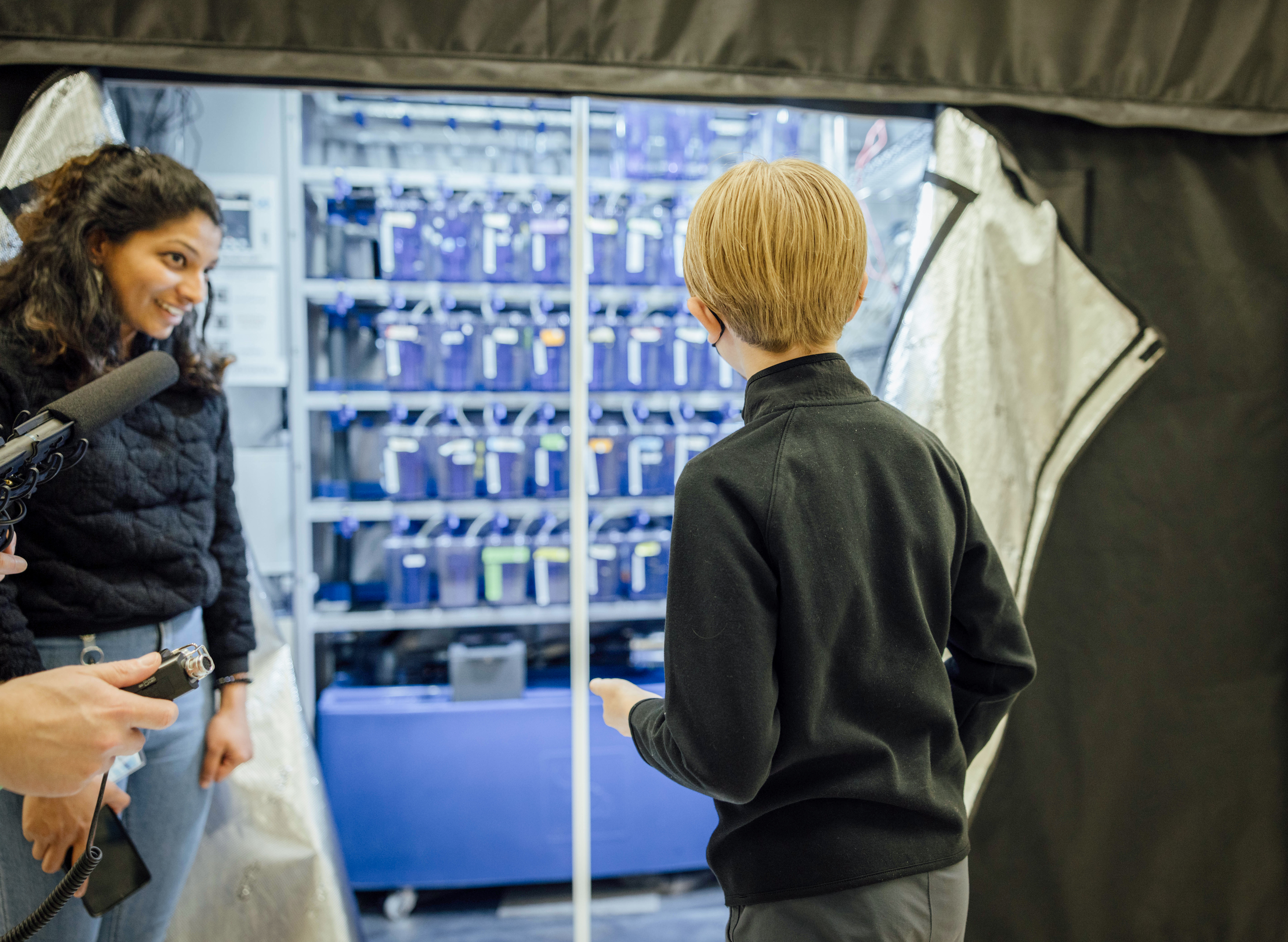
Nate: So we’re moving into the wet lab and there’s this big black tent and they’re about to unzip it. But above there’s this sign. Oh, the zebrafish hotel. The sign says Le Petit Hotel de Poisson. The small hotel of fish, I believe. And here in this majestic black zip up tent is where all of the zebrafish live. OK, so in front of me is the zebrafish hotel and there are a bunch of tanks, each filled with a bunch of different zebrafish. Some of them look fairly normal, but others of them are red tinted or fairly red looking.
Merlin Lange: It’s because there’s so much like red fluorescence inserted in the genome that even like the pigments are becoming red. Wow. And we also have those fish here on the bottom, right here.
Nate: Oh yeah, those look pretty white. They’re completely see through. And it’s it’s all very cool looking. Every tank in Le Petit Hotel was home to a different type of genetically modified zebrafish.
Shruthi VijayKumar: So each of these tanks, they’re basically a different transgenic line and they express a different fluorescence gene.
Nate: What, first off, what is a transgenic line?
Shruthi VijayKumar: So basically a transgenic line is if you want to enhance a particular gene or if you want to study a particular organism, you use genetic modification like CRISPR-Cas9, which is a gene modification method where you can, you know, either delete a particular gene of interest or put in a gene which you want to see. For example, in case you want to study spinal cord development, you can put in GFP, which is a fluorescence which enhances the spinal cord. So your spinal cord will fluoresce under the microscope. So this will allow you to see the development of the spinal cord under the microscope. So it helps you see any organ you want technically. So you can, let’s say, put a red color signal on the heart or a green color signal in the eye and you can see this develop. So basically each of these is a transgenic line. You create a line which allows you to see these organs.
Sandy Schmid: Nate, you heard Manu is putting these fluorescent proteins in at the cellular level. Yeah. You can also do it at the level of a whole zebrafish.
Nate: This is Sandy Schmid. You’ll remember that she’s the chief scientific officer at the Biohub.
Sandy Schmid: So whereas Manu was lighting up individual proteins by lighting up proteins that belong in individual organs, like your brain or your heart or your kidney, you can light up the fish. So it’s the same technology now applied at the whole organism level as opposed to at the single cell level.
Nate: The fluorescent proteins or genetic glow sticks that we talked about with Manu in the last episode are called GFP, which stands for green fluorescent proteins. The discovery of GFP won a Nobel Prize in 2008. It’s allowed scientists to see things that they just couldn’t see before.
Sandy Schmid: All of a sudden you can watch these proteins, these tissues, these animals in real time under the microscope and you couldn’t do that before you could use these fluorescent proteins. So that was a pretty good Nobel Prize.
Nate: So now you can see all of the different things under the microscope that you wouldn’t have been able to before. And you can actually see the individual cells and the proteins and you can map them and do a bunch of different things that you wouldn’t have been able to before that.
Sandy Schmid: Absolutely. The cell biologists before GFP only looked at dead cells. Right. And now we can look at living cells and they only look at dead embryos at different time points. Right. And now you can look at a living embryo and watch the whole process.
Nate: Watching the entire development process unfold under a microscope is what happens after creating those different transgenic lines of zebrafish. And this is where things start getting really exciting.
Merlin Lange: So now we can go to see actually under a microscope what our fluorescence or transgenic zebrafish looks like. Right. So we are entering in the microscopy area.
Nate: Oh, all right.
Merlin Lange: So why the microscopy area is different from the other part of the wet lab is because we need like total darkness. So those are like rooms with curtains that prevent any light to enter in.
Nate: I see. Okay.
Merlin Lange: So right now this is the sample mounting room.
So we have two pretty basic microscopes.
Nate: They’re called binocular microscopes because they have two eyepieces instead of one. This means that you can look through these microscopes with both eyes.
Merlin Lange: So it’s the microscope that we are using to mount the sample that then will go in the light sheet microscope. And this one is mainly for screening. And we basically, what we can see under this is those two zebrafish. You want to look at them under the binocular?
Nate: Yeah, that would be nice.
Merlin Lange: Can you see something?
Nate: Yes, I can. Oh, yep. Those are embryos. That’s a feisty little guy.
Merlin Lange: Are they moving?
Nate: Yeah, the one was squirming around a little.
Merlin Lange: Yeah, they are relatively late in development. So they are like post 24 hours. After fertilization, we usually study under the microscope zebrafish from around 5 hours post fertilization to 24 hours post fertilization.
Nate: I see. But yes, those guys are pretty far along in development. They’re starting to move around and squirm a little. But yeah, they’re still very red. They have little eyes. Yeah, they look like little fish eggs.
Merlin Lange: And can you see the pigments here?
Nate: Yeah, there is pigment.
Merlin Lange: I was mentioning about the transgenic line that has no pigment.
Nate: Because that’s not the light reflecting off the mirror. That is their pigment, right?
Merlin Lange: Yes, yes, yes. So basically, if we want to image with the pigment, it would be very difficult. So it’s why it’s very useful to have transgenic fish that have no pigment at all. Because you can then do imaging there.
Nate: Because these ones are bright red.
Merlin Lange: Yes, very bright.
Nate: Very cute too.
Merlin Lange: So now let’s have a look at the light sheet microscope.
Nate: Alright, sounds good.
Sandy Schmid: Are you going to show a microscope that doesn’t look anything like a microscope?
Nate: Oh, alright.
We headed into the room next door where they kept the light sheet microscope. Yep, that doesn’t look like a microscope.
Loïc Royer: So what we’re seeing here is essentially our first simultaneous relative view light sheet microscope. So that’s the first microscope we built here on my team.
Nate: This looks like somebody trying to create like a death laser in a movie or something.
Loïc Royer: Pretty much. Guess what? There’s actually a laser in the system right now.
Nate: Yeah?
But yeah, essentially we surround the sample with microscope objectives, which are these things here that you see here. I don’t know if you can…
Nate: And so the microscope objectives that Loïc is talking about are basically the microscope’s lenses. And these lenses are positioned around the sample in a way that allows you to take 3D images of it.
Loïc Royer: And we can do that every 30 seconds. So we get a 3D image of the full embryo every 30 seconds, which lets us essentially get a movie, a three-dimensional movie of the whole process of development from almost from a single cell all the way to a fully functional organism.
Nate: Loïc pulled up some of these movies for us so that we can see the development in action.
Loïc Royer: So here is the head here.
Nate: Yeah.
Loïc Royer: And then the tail. And then what’s happening is that you basically we are segmenting the cells, which means we are identifying each nucleus. And there was a different color because we could essentially count them and then identify them. We’re rewinding the video. And now what we’re doing is we’re tracking all these nuclei. So you see the little tracks we follow because we really know where they are in the computer. We can produce the data set that tells us all the coordinates of all these cells over time. And so we can really follow them and understand where they’re going, where they’re coming from, and eventually the cells divide. So then we also want to understand the relationship between the cells and their parents and their daughter cells. And now we’re going to follow one cell. So we keep following the cell. We follow it. We follow it. Imagine it’s just one cell and there’s so many more cells around it. And it’s very difficult to follow it because they all look the same, right? But we actually have very, very good algorithms that allow us to follow this cell very accurately. And we keep following that single cell and poof, it’s split in two. Did you see that? And the cells, the cells eventually divide.
Nate: Yeah.
As amazing as these videos are, we actually still don’t know what’s going on in them. It’s a mystery. But it’s a mystery that the people at the Chan Zuckerberg Biohub are working very hard to solve.
Loïc Royer: The truth is that we don’t know what really happens here. Each cell is as complex as perhaps any of the robots we’ve ever built as humans. They actually have little programs telling them what to do. They’re following certain trajectories. They’re talking to their neighbors, figuring out who they are, what they should be doing, how they should organize themselves. Super complex stuff.
Nate: Super complex, but also super cool to watch. We have some of those videos linked in the description and trust me, you’re gonna want to see them. On the next episode, we’re going to be concluding this four-part series with a discussion between myself, Stephen Quake, who is the head of science at the Chan Zuckerberg Initiative, and Priscilla Chan, who is the co-founder and co-CEO of CZI. You’re not going to want to miss it. So stay tuned for next time.
There you have it, folks. The Show About Science is complete. Special thank you to Loïc, Merlin, Shruti, and Sandy for taking me on a wonderful tour of Le Petit Hotel de Poisson. This episode wouldn’t have been possible without everyone at CZI and the CZ Biohub. Extra special thanks to Patricia Condon, Pete Farley, Jeff McGregor, Dale Ramos, and Sandy Schmid. And our theme music, as always, was written by Jeff, Dan, and Theresa Brooks. Okay, Dad, you can shut the recording off.
Leave a Reply